 A very simple case. Oral mucosal biopsy for a “suspect pigmented lesion”.
The sections show mucosal hyperplasia; no dysplasia or melanocytic proliferation.
In the superficial stroma there is an interstitial granular black pigment; taken up by macrophages, also present in blood vessel walls.
——————————————————————————AMALGAM TATTOO.——————————————————————————
J Dermatol. 2011 Jan;38(1):101-3. doi: 10.1111/j.1346-8138.2010.01007.x. Epub 2010 Sep 20.
Amalgam tattoo of the oral mucosa mimics malignant melanoma.
Amano H, Tamura A, Yasuda M, Yamanaka M, Takeuchi Y, Sasaoka K, Yokoo S, Ishikawa O.
——————————————————————–
Laryngorhinootologie. 1991 Sep;70(9):515-7.
[Amalgam tattoo--an important differential diagnosis from malignant melanoma of the mouth mucosa].
[Article in German]
Tolsdorff P, Schützenberger KH.
Arzt für HNO-Krankheiten und Plastische Operationen, Bad Honnef.
Abstract
Amalgam tattoos develop as an amalgam deposit often as a result of continuous contact between an amalgam filling and the gingiva or amalgam fragments embedded in the oral tissue during dental filling or surgical operations. Sometimes fragments of amalgam restorations are broken off during extraction and embed in the adjacent soft tissue. In our case a small piece of amalgam had broken off during an extraction after retrograde filling up this tooth some years before following a resection of the dental root. Embedded in the depth of the bony resection cavity the piece of amalgam had produced the amalgam tattoo 13 years later.
———————————————————————-
Br J Oral Maxillofac Surg. 2009 Jun;47(4):313-5. Epub 2009 Feb 28.
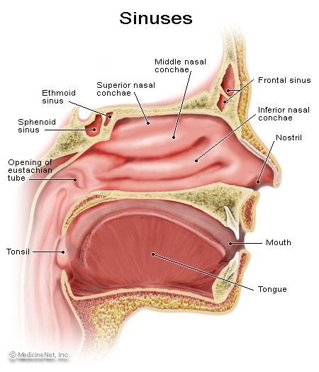
Aspergillosis of the maxillary sinus secondary to a foreign body (amalgam) in the maxillary antrum.
Burnham R, Bridle C.
Oral and Maxillofacial Surgery Centre, the Royal London Hospital, London, United Kingdom.
Comment in:
Br J Oral Maxillofac Surg. 2010 Jan;48(1):64; author reply 64-5.
Abstract
We report a case of a Maxillary sinus aspergilloma, which presented after 2 years of symptoms of chronic sinusitis. there was an isolatable triggering event of extrusion of an amalgam filling material into the sinus. this was a complication of surgical extraction of the upper right second molar by his general dental practitioner.
———————————————————————-
Ultrastruct Pathol. 2005 Sep-Oct;29(5):405-13.
Chronic inflammation and pain inside the mandibular jaw and a 10-year forgotten amalgam filling in an alveolar cavity of an extracted molar tooth.
Kaufmann T, Bloch C, Schmidt W, Jonas L.
Department of Maxillofacial Surgery, Medical Faculty, University of Rostock, Rostock, Germany.
Abstract
A 55-year-old woman, suffered from severe pain in her mandibular jaw for several years. A metallic artifact of about 2(3) mm was detected by a panorama radiography in an edentulous region with a surrounding inflammation in close contact to the canal of the mandibular nerve. Inflammated tissue with the central metallic inclusion was removed from the bone under local anesthesia and operation. Postoperatively, pain and missensitivity disappeared within 1 week. although the patient had no macroscopically visible so-called amalgam tattoo, the metallic cube was identified as amalgam by the detection of mercury, silver, tin, copper, and zinc using energy dispersive X-ray analysis (EDX) in a scanning electron microscope (SEM). nevertheless, brown to black pigments in the connective tissue matrix and inside histiocytes, fibroblasts, and multinucleated foreign giant cells of the surrounding inflammatory tissue were observed by light and electron microscopy. however, the elemental analysis by EDX in SEM or by electron energy loss spectroscopy in transmission electron microscope detected only silver, tin, and sulfur but no mercury in these precipitates and in the residual bodies of phagocytes. the presented case demonstrates a seldom complication of amalgam deposition in the tissue. the authors assume that the chronic pain results from a forgotten amalgam filling inside an alveole after extraction of a molar tooth, causing a chronic inflammation by resolving mercury and other toxic elements out of the metallic artifact.
———————————————————————-
J Occup Med Toxicol. 2011 Jan 13;6(1):2.
Is dental amalgam safe for humans? the opinion of the scientific committee of the European Commission.
Mutter J.
Department of Environmental and integrative medicine Lohnerhofstraße 2, 78467 Constance/Germany. .
Abstract
ABSTRACT: It was claimed by the Scientific Committee on Emerging and Newly Identified Health Risks (SCENIHR)) in a report to the EU-Commission that “….no risks of adverse systemic effects exist and the current use of dental amalgam does not pose a risk of systemic disease…” [1, available from: ec.europa.eu/health/ph_risk/committees/04_scenihr/docs/scenihr_o_016.pdf].SCENIHR disregarded the toxicology of mercury and did not include most important scientific studies in their review. But the real scientific data show that:(a) Dental amalgam is by far the main source of human total mercury body burden. this is proven by autopsy studies which found 2-12 times more mercury in body tissues of individuals with dental amalgam. Autopsy studies are the most valuable and most important studies for examining the amalgam-caused mercury body burden.(b) these autopsy studies have shown consistently that many individuals with amalgam have toxic levels of mercury in their brains or kidneys.(c) there is no correlation between mercury levels in blood or urine, and the levels in body tissues or the severity of clinical symptoms. SCENIHR only relied on levels in urine or blood.(d) the half-life of mercury in the brain can last from several years to decades, thus mercury accumulates over time of amalgam exposure in body tissues to toxic levels. however, SCENIHR state that the half-life of mercury in the body is only “20-90 days”.(e) Mercury vapor is about ten times more toxic than lead on human neurons and with synergistic toxicity to other metals.(f) most studies cited by SCENIHR which conclude that amalgam fillings are safe have severe methodical flaws.
———————————————————————-
Crit Rev Toxicol. 2007;37(6):537-49; discussion 551-2.
Comments on the article “the toxicology of mercury and its chemical compounds” by Clarkson and Magos (2006).
Mutter J, Naumann J, Guethlin C.
University Hospital, Institute for Environmental Medicine and Hospital Epidemiology, Freiburg, Germany.
Comment on:
Crit Rev Toxicol. 2006 Sep;36(8):609-62.
Abstract
Clarkson and Magos (2006) provide their perspectives on the toxicology of mercury vapor and dental amalgam. as scientists who are involved in preparing a German federal guideline regarding dental amalgam, we welcome additional scientific data on this issue. however, Clarkson and Magos do not present all the relevant studies in their review. the additional data provided here show that: (a) Dental amalgam is the main source of human total mercury body burden, because individuals with amalgam have 2-12 times more mercury in their body tissues compared to individuals without amalgam; (b) there is not necessarily a correlation between mercury levels in blood, urine, or hair and in body tissues, and none of the parameters correlate with severity of symptoms; (c) the half-life of mercury deposits in brain and bone tissues could last from several years to decades, and thus mercury accumulates over time of exposure; (d) mercury, in particular mercury vapor, is known to be the most toxic nonradioactive element, and is toxic even in very low doses, and (e) some studies which conclude that amalgam fillings are safe for human beings have important methodogical flaws. therefore, they have no value for assessing the safety of amalgam.